A mammogram is an X-ray of the breast that’s used to screen for and diagnose breast cancer. 3D mammography (known as Tomosynthesis) is the most modern screening and diagnostic tool available for early detection of breast cancer. While conventional methods generate flat images, Tomosynthesis creates a detailed three-dimensional depiction of the breast. This enables greater accuracy, earlier breast cancer detection and decreased recall rates – meaning that fewer women are brought back for additional screening. It’s relatively new technology that’s growing in popularity around the world.
Tomosynthesis takes multiple X-rays at different angles to create a 3D picture of the breast. It uses relatively low radiation doses and is therefore considered safer than traditional 2D breast imaging methods. However, 3D imaging is sometimes complemented by a 2D perspective and often both types are prescribed to gain a full understanding of the issue. Merging both techniques has been found to increase the number of cancers detected. The 3D mammogram machine at Durdans Hospital can generate both 2D and 3D images. This reduces the need for follow-up imaging and can save time and money.
The technology is used to look for breast cancer in people who have no outward signs or symptoms. It can also be used to identify the cause of breast pain, nipple discharge and investigate suspicious lumps and thickening. It’s particularly useful for detecting cancer in dense breast tissue – dense breasts have greater amounts of connective tissue and are difficult to visualise via traditional mammograms (because both dense tissue and tumours appear white on the image). Tomosynthesis enables doctors to see beyond the areas of density and identify tumours that may not have been visible otherwise. The clearer images generated from 3D mammograms are thought to help detect 41% more invasive cancers and significantly decrease the number of recalls for additional imaging.
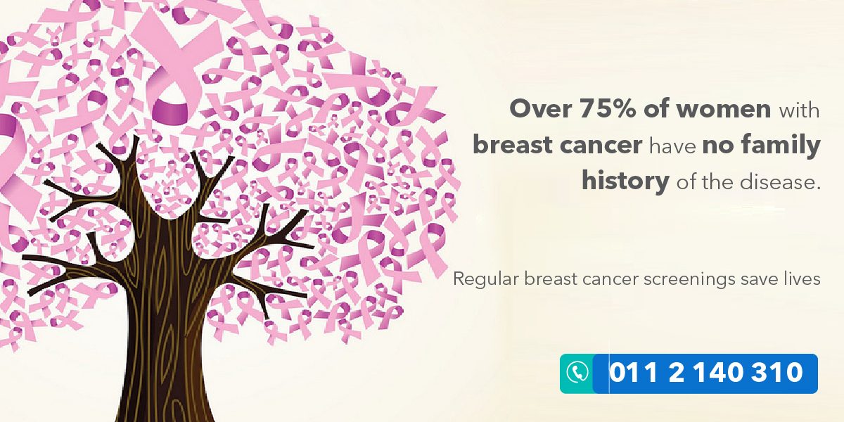
Mammograms are recommended every 1 to 2 years for women over 40 years or for younger women with a genetic predisposition for breast cancer. You don’t need to experience any signs or symptoms of a breast abnormality to be considered. 80% of tumours found during a mammogram are benign (not cancerous). Although false alarms can be stressful, regular screenings help with early detection – early detection saves lives. Eight out of ten women diagnosed with breast cancer have no family history of the disease. Mammography detects most cancers in women who show no symptoms or who are considered low risk.
A mammogram is an important step to taking care of your health but not knowing what to expect can be stressful. During your exam, your breast will be momentarily compressed against X-ray plates and an X-ray arm will sweep over your breast in an arc and take multiple images in a matter of seconds. A computer will then assemble a 3D image of your breast tissue. This can either be analysed as a whole or examined in small fractions in greater detail. Metallic particles in deodorants, powders, creams and perfumes can interfere with the imaging process, so don’t use them under your arms or on your breasts before your exam.
The Durdans Hospital radiology team is available 24/7 to provide convenient and high-quality imaging services. Call 011 2 140 310 to book your appointment today
