Dr Manohari Seneviratne
Consultant Physician
Dr Chaminda Garusinghe
Consultant Diabetologist & Endocrinologist
Osteoporosis is defined as a disease characterised by low bone mass and micro-architectural deterioration of bone tissue leading to enhanced bone fragility and an increase in fracture risk
Osteoporosis is a condition affecting mainly the elderly causing brittle bones resulting in fractures even at minor trauma. Hip, vertebrae and distal forearm are the commonest sites affected due to this disease causing a great deal of disability and even death. It has been found that 3 month and 1 year survival following a hip fracture is fairly low.
Osteoporosis is defined as a disease characterised by low bone mass and micro-architectural deterioration of bone tissue leading to enhanced bone fragility and an increase in fracture risk. Osteoporosis results from increased bone breakdown by osteoclasts and decreased bone formation by osteoblasts leading to bone loss.
Common risk factors
- Increasing age
- Genetics- a family history of osteoporosis or low trauma fractures (Colles fracture, vertebral, hip, proximal humerus
- Early menopause (less than 40 years)
- Low body weight
- Prolonged steroid use (glucocorticoids > than 7.5 mg more than 3 months)
- Calcium and vitamin D deficiency
- X-rays showing osteopenia
Osteoporosis is clinically silent until the onset of a facture. 2/3 of vertebral fractures are not seen by doctors. Typical signs of a vertebral fracture –a sudden episode of well-localised pain which may not be related to injury that radiates in a girdle fashion. Osteoporosis does not cause generalised skeletal pain most of the time.
WHO classified osteoporosis based on the bone mineral density as detected in DXA scan. The patient’s bone density is compared with that of sex and ethnicity matched healthy young man or a woman. The subject’s bone density is expressed in terms of standard deviations. WHO guidelines for diagnosis of postmenopausal osteoporosis are as follows:
Investigate to exclude an underlying cause
DXA scan can only tell us that the bone mass is reduced. Even though this is mostly to do with post-menopausal osteoporosis, there are so many other causes to be excluded before treatment. As multiple myeloma is an important secondary cause in the affected age group, Full blood count, ESR are very important screening tests. . Liver function test will be abnormal with excess alcohol use and cirrhosis which are also risk factors for osteoporosis. Thyroid functions should be done to exclude clinical as well as subclinical thyrotoxicosis causing osteoporosis. Vitamin D level are generally low in Sri Lankans due to nutritional deficiency and low sun exposure and the presence of dark skin.
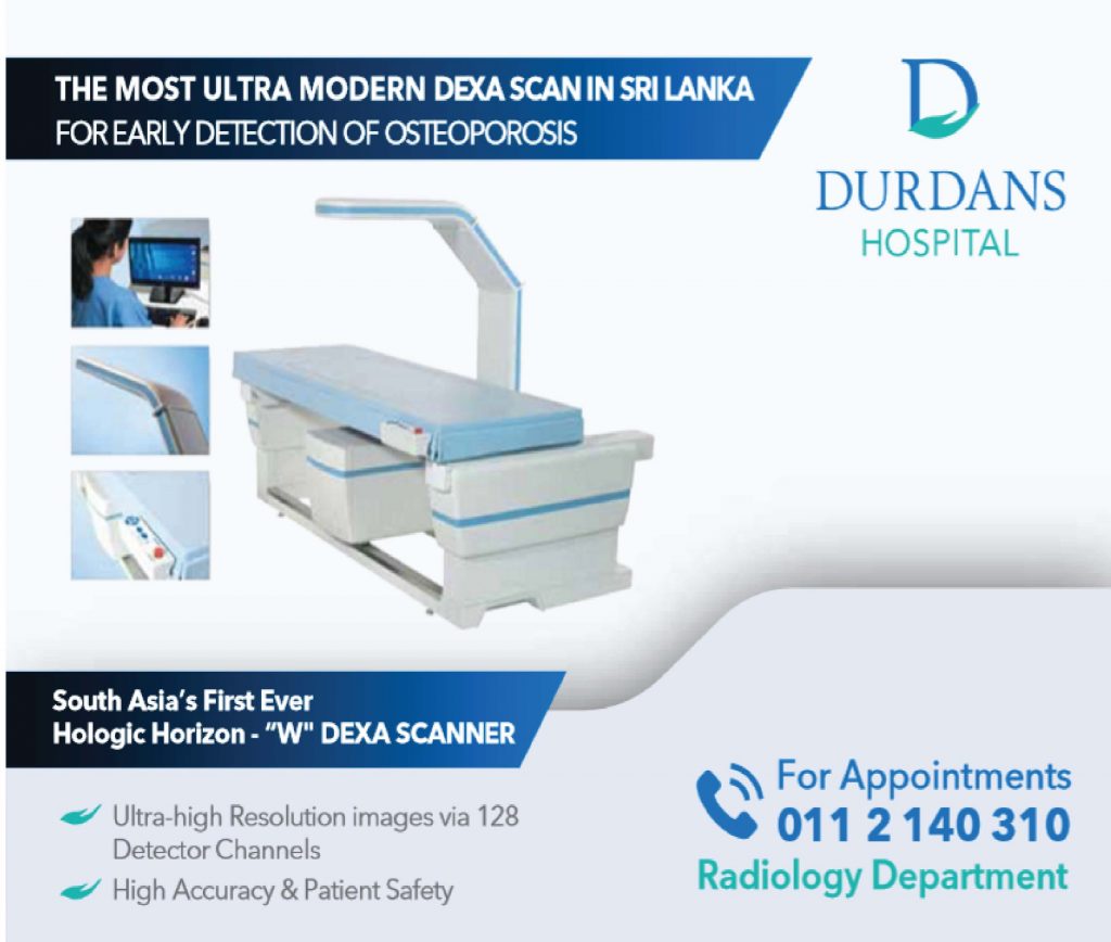
DEXA is the definitive way to diagnose osteoporosis. It should be done for patients at risk or with osteopenia on X- rays. The DEXA will have to be repeated to see response to treatment.
Treatment
All those who are affected will have to take be on calcium and Vitamin D supplements. Achieving a higher peak bone mass in young age by good nutrition as well as physical activities should be encouraged for younger people.
Alendronate 70mg once a week or Zolindronic acid 5 mg IV slow infusion over half an hour once yearly are the most popular modes of treatment. Alendronate must be taken with a full glass of water on an empty stomach and the patient must keep his body upright for 30 minutes till the drug passes thought the gastro duodenal junction. This is important to avoid gastric irritation which can be severe with Bisphosphonates. Zolundronate is known to cause osteonecrosis of the jaw especially with poor dental hygiene therefore it is good to attend to the patient’s dental hygiene before commencing treatment. Atypical fractures of the femur have also been reported and now it is recommended to give a drug holiday for one year after 5 years of treatment.
Primary prevention, case detection with DXA scan and proper treatment of osteoporosis can greatly reduce the morbidity and mortality of osteoporosis.


